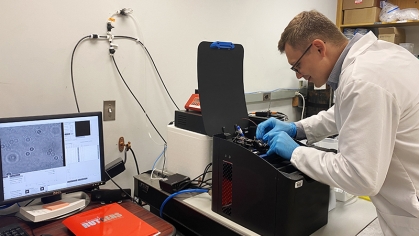
LAB REPORT: Developing Optical Imaging Systems to Diagnose and Manage Cancer and Other Diseases
As an undergraduate at the University of Manchester in England, Department of Biomedical Engineering (BME) Professor Mark Pierce majored in physics before exploring surgical lasers while earning his PhD.
"That helped me see how physics can have applications in human health so after grad school, I joined a lab working on using lasers for imaging," he recalls. "Light has many properties we can use for diagnosis and treatment of disease, so there were many directions to explore."
Since joining the BME department in 2011, Pierce's lab has launched fruitful—and frequent federal, state, and industry funded—explorations of many of these avenues, while maintaining a sharp research focus on developing optical imaging systems to aid in cancer and other disease diagnoses and management.
Seeing Cells and Tissues in a New Light
"My lab works on developing optical imaging systems aiming to give researchers and clinicians information about cells and tissues, beyond what they can see with the naked eye," Pierce explains.
"Light transmitted through or reflected from tissue carries information about that tissue, which we can analyze to assess health," he notes. "Depending on the application, sometimes we zoom in to look at the cellular or microstructural level, sometimes we zoom out to look at an entire organ."
In his lab, Pierce—with the ongoing involvement of graduate and undergraduate students—has continued to explore the use of wavelengths, or colors, in the short-wave infrared spectral region. He explains, "Most researchers have focused on using visible and near-infrared light, but I've been imaging and treating tissue with these wavelengths since graduate school. In recent years, more people are exploring this spectral region, and more companies are providing compatible instruments and components."
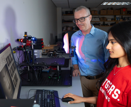
By leveraging AI advances, the lab is developing deep learning models that can quickly analyze imaging data and estimate tissue properties in real time. "The inference speed of these models is valuable when each pixel in an image needs to be analyzed separately," he adds.
A current focus of the lab is seeing if optical imaging can measure and assess skin hydration and sweat dynamics. "My recent PhD student Tommy Livecchi showed that our skin properties change when we sweat during exercise and these changes can be measured with a specialized camera system," Pierce notes, while another study used thermal imaging to see sweat pores being activated by exercise.
Other potential applications for skin hydration measurements include assessment for dermatologic skin conditions such as eczema and psoriasis, which are related to skin's barrier function. "If this barrier is damaged, the skin loses water and becomes hydrated, increasing the risk of developing these conditions," he explains.
Developers of cosmetic products such as moisturizers would benefit from being able to measure how well a product is maintaining superficial skin hydration. "Such measurements would help them modify their formulations and inform their marketing claims about how well their products work," Pierce adds.
Putting Discoveries to Work
Over time, clinicians have used a number of Pierce's innovations, ranging from therapeutic lasers for breaking down kidney stones and for dentistry to optical coherence tomography, or OCT. "I continue to work on this non-invasive imaging method which is used to diagnose conditions in the retina and cardiovascular systems," he reports.
According to Pierce, optical imaging—like other medical imaging modalities—can work together with a contrast agent to highlight specific tissues of interest. He has collaborated with other School of Engineering colleagues—including form BME professor Prabhas Moghe; department chair Charles Roth; and Department of Materials Science and Engineering Professor Richard Riman—to develop a nanoparticle contrast agent able to label cancerous tumors within the body and emit infrared light for imaging.
The group additionally designed and built an instrument that enables the imaging of the nanoparticle contrast agent. "Without the imaging system, we wouldn't have been able to demonstrate the capabilities of the contrast agent."
Nanolink Imaging, a start-up company, was established to commercialize this innovative technology and, Pierce reports, is currently working on putting the first product on the market.